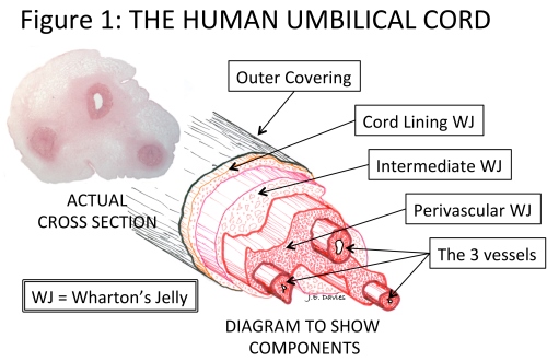
Describe the Umbilical Cord Blood Vessels
The blood in the arteries contains waste products such as carbon dioxide from the babys metabolism. The umbilical cords ___ umbilical artery and ___ umbilical vein are surrounded by.
Umbilical cord development begins in the embryologic period around week 3 with the formation of the connecting stalk.

. What blood vessels are located within the umbilical cord. This enriched blood flows through the umbilical vein toward the babys liver. 2 1 Whartons jelly.
Pair of vessels that runs within the umbilical cord and carries fetal blood low in oxygen and high in waste to the placenta for exchange with maternal blood umbilical vein. A small greenish structure on the underside of the liver. The afterbirth is a placenta with part of the umbilical cord attached.
When the cord lies below presenting part. Click to see full answer. A normal cord has two arteries small round vessels with thick walls and one vein a wide thin-walled vessel that usually looks flat after clamping.
The objective is to describe the duration and patterns of blood flow through the umbilical vessels during DCC. It is enclosed inside a tubular sheath of amnion and consists of two paired umbilical arteries and one umbilical vein. The researchers inserted beads with a diameter of 05 µm into blood vessels in the placenta.
Oxygen and nutrients from the mothers blood are transferred across the placenta to the fetus through the umbilical cord. 5 rows The blood inside the umbilical cord is called cord blood and it contains undifferentiated. Describe what happened to the duct from the gallbladder.
Did it join other ducts. The placenta is an organ that connects the developing fetus to the uterine wall. There are two umbilical veins and two umbilical arteries in the cord.
However some babies have just one artery and vein. It has been shown that this blood contains at least three populations of stem cells each with uni-que. After birth the distal part of the artery obliterates and becomes the medial umbilical ligamentThe proximal part of the artery still remains functional providing a blood.
Typically an umbilical cord has two arteries and one vein. Occasionally only two vessels one vein and one artery are present in the umbilical cord. Single vessel that originates in the placenta and runs within the umbilical cord carrying oxygen- and nutrient-rich blood to the fetal heart.
A study was done on donated afterbirths. The umbilical cord which connects your baby to the placenta contains three vessels. Waste products and carbon dioxide from the fetus are sent back through the umbilical cord and placenta to the mothers circulation to be eliminated.
Blood vessels of the allantois become the umbilical blood vessels. The purpose of the study was to find the maximum size of particles that can pass through the placenta and enter the umbilical cord. Two arteries which carry blood from the baby to the placenta and one vein which carries blood back to the baby.
Where did the ducts end. Umbilical vein vena umbilicalis The umbilical vein is an important part of the fetal circulationUnlike regular veins in adulthood the fetal umbilical vein carries oxygenated blood from the placenta into the growing fetus. This condition is known as a two-vessel cord diagnosis.
Describe prolapse of the cord. Two smaller umbilical arteries and one larger umbilical vein. Blood flow in the unborn baby follows this pathway.
Statements a and b are both correct. Through the blood vessels in the umbilical cord the fetus receives all the necessary nutrition oxygen and life support from the mother through the placenta. Thrombosis of Umbilical Cord is a condition in which one of the blood vessels of the umbilical cord gets blocked or obstructed.
A body stalk that connects the embryo to the chorion becomes the umbilical cord. How many blood vessels are in the umbilical cord of a fetal pig. Delayed umbilical cord clamping DCC affects the cardiopulmonary transition and blood volume in neonates immediately after birth.
By week 7 the umbilical cord has fully formed composed of the connecting stalk vitelline duct and umbilical vessels surrounding the amniotic membrane. It is a disc shaped reddish brown structure that connects the fetus to. However little is known of blood flow in the umbilical vessels immediately after birth during DCC.
This can result in life-threatening bleeding in the baby. A thrombosis indicates the clotting of blood in a blood vessel. The umbilical cord has three blood vessels one for oxygen and nutrients from the placenta to the baby and two arteries carry deoxygenated fetal blood from the.
The umbilical cord houses blood vessels that join the embryo to the placenta In the placenta the blood vessels from the umbilical cord run close to the mothers blood vessels but arent actually connected to them The close proximity of the blood vessels allows nutrients oxygen vitamins and waste products to be exchanged between mother and embryo The head develops before. It contains one vein which carries oxygenated nutrient-rich blood to the fetus and two arteries that carry deoxygenated nutrient-depleted blood away. There it moves through a shunt called the ductus venosus.
Umbilical artery Arteria umbilicalis The umbilical artery is a paired vessel that arises from the internal iliac arteryDuring the prenatal development of the fetus it is a major part of the fetal circulation. Umbilical cord blood is the blood found in the vessels of the umbilical cord and placenta. One of the umbilical arteries is visible protruding from the cut edge.
The blood vessels unprotected by the Whartons jelly in the umbilical cord or the tissue in the placenta sometimes tear when the cervix dilates or the membranes rupture. The cord is plump and pale yellow in appearance. The umbilical cord is the vital connection between the fetus and the placenta.
What makes a false knot look like a knot in the cord. A large artery that carries blood from the left. During fetal life the umbilical vein arises within the placenta and passes through the umbilical cord along with the paired umbilical arteries.
Which statements accurately describe the umbilical cord. This infant is 7 hours old. The umbilical cord is a bundle of blood vessels that develops during the early stages of embryological development.

Umbilical Cord Structure Functions Storage Abnormalities Infections


Comments
Post a Comment